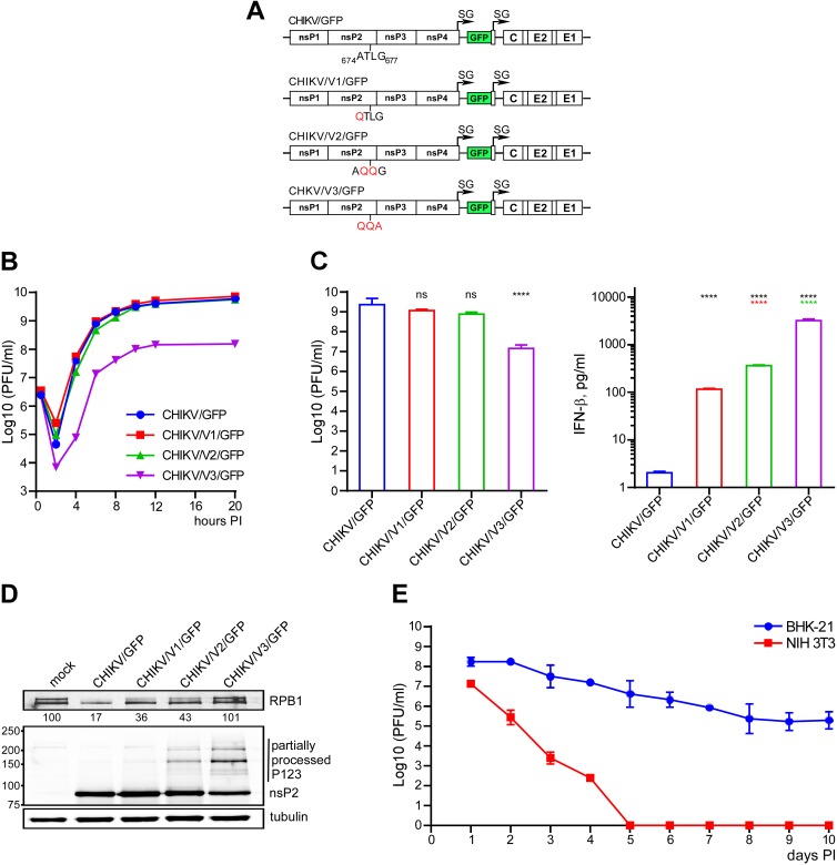
FIG 2.
SINV-specific mutations in CHIKV-specific VLoop differentially affect viral replication rates and activation of the antiviral response. (A) Schematic presentation of recombinant CHIKV genomes encoding indicated peptides. (B) NIH 3T3 cells were infected with the indicated viruses at an MOI of 20 PFU/cell. Media were replaced at the indicated times p.i., and viral titers were determined by plaque assay on BHK-21 cells. (C) NIH 3T3 cells were infected with control and mutant viruses at an MOI of 20 PFU/cell. Viruses were harvested at 18 h p.i. Infectious titers and concentrations of the released IFN-β were determined in the same samples as described in Materials and Methods. The significances of differences in titers and IFN-β concentrations were estimated by one-way analysis of variance (ANOVA; n = 3). ****, P < 0.0001. (D) NIH 3T3 cells were infected with the indicated viruses at an MOI of 20 PFU/cell and harvested at 8 h p.i. The lysates were analyzed by Western blotting for the levels of RPB1 and for the ns polyprotein processing using RPB1-, nsP2-, and tubulin-specific Abs and corresponding infrared dye-labeled secondary Abs. Membranes were scanned on Odyssey imager (LI-COR). (E) BHK-21 and NIH 3T3 cells were infected with CHIKV/V3/GFP at an MOI of 10 PFU/cell and then incubated for 10 days. Media were changed every 24 h, and cells were split upon reaching confluency. Viral titers were determined by plaque assay on BHK-21 cells. Data are presented as means ± SDs from two independent experiments.

