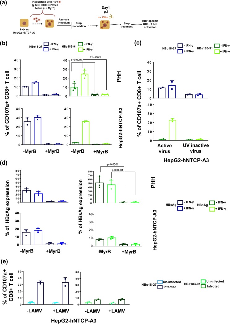
FIG 5.
HBV epitope presentation requires NTCP-mediated infection and viral protein synthesis. (a) Schematic representation of infection of HepG2-hNTCP-A3 cells and PHH in the presence or absence of 800 nM Myrcludex B (MyrB). (b) Frequency of HBV-specific CD8+ T cells expressing CD107a among total CD8+ T cells after 5 h of coculture (E/T ratio of 1:2) with infected PHH or HepG2-hNTCP-A3 cells at day 1 postinfection. Targets were infected in the presence or absence of MyrB. In addition, cells were either treated with IFN-γ or left untreated as described in the legend of Fig. 4. (c) Ability of CD8+ T cells specific for HBc18-27 or HBs183-91 to recognize HepG2-hNTCP-A3 cells infected with HBV or UV-inactivated HBV in the presence or absence of IFN-γ. Bars show CD8+ T cells positive for CD107a among total CD8+ T cells after 5 h of coculture with target cells at day 1 postinfection (E/T ratio of 1:2). Data shown as means ± standard deviations of two independent experiments. (d) Expression of HBcAg and HBsAg in the presence or absence of MyrB treatment in both PHH and HepG2-hNTCP-A3 infected targets at day 1 p.i. is shown. Targets were either treated with IFN-γ (bright shades) or untreated (dark shades). (e) Frequency of CD107a-positive CD8+ T cells specific for HBc18-27 and HBs183-91, cocultured with HBV infected (dark shades) or uninfected (bright shades) HepG2-hNTCP-A3 cells treated or untreated with 10 μM lamivudine. Data are from day 3 postinfection.

