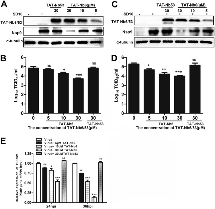
FIG 6.
Inhibition of PRRSV SD16 infection and replication by TAT-NB6 in PAMs. PAMs were infected with SD16 at an MOI of 0.01 for 1 h, and then the cell culture media were replaced with fresh RPMI 1640 containing 3% FBS and TAT-Nbs at the indicated concentrations. TAT-Nbs and PRRSV Nsp9 were detected at 24 hpi (A) and 36 hpi (C) by Western blotting using anti-His MAb and mouse anti-Nsp9 antiserum, respectively. Progeny virus released in the cell medium was measured by TCID50 at 24 hpi (B) and 36 hpi (D). (E) Relative levels of PRRSV RNA were detected by RT-qPCR using PRRSV Nsp9-specific primers. The GAPDH mRNA level served as an internal reference. Data are expressed as means ± SD from three independent experiments. P values were calculated using ANOVA as <0.05 (*), <0.01 (**), and <0.001 (***) compared with cells infected with PRRSV alone (ns, not significant).

