FIG 4.
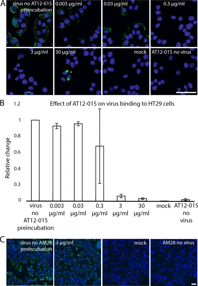
Antibody AT12-015 blocks virus binding to HT29 cells. (A) Representative fluorescence images of HPeV3 incubated in the presence or absence of various amounts of AT12-015 antibody. Cell nuclei were visualized using a Hoechst stain (blue), and bound virus was scored by measuring Alexa Fluor 488 intensity (green). Pictures were acquired using a 20× objective. Scale bar, 50 μm. (B) Effect of preincubation of HPeV3 with different amounts of human monoclonal antibody AT12-015. The results are the averages from three repeats of the cold binding assay. The error bars represent the standard errors of the means (SEMs). (C) AM28 had no effect on HPeV3 binding to HT29 cells. Representative fluorescence images of HPeV3 incubated in the presence (3 μg/ml) or absence (virus no AM28 preincubation) of AM28 antibody and added to the cells for binding. Noninfected cells (mock and AM28 no virus) served as the controls. Stained as in panel A and visualized with a 10× objective. Scale bar, 50 μm.

