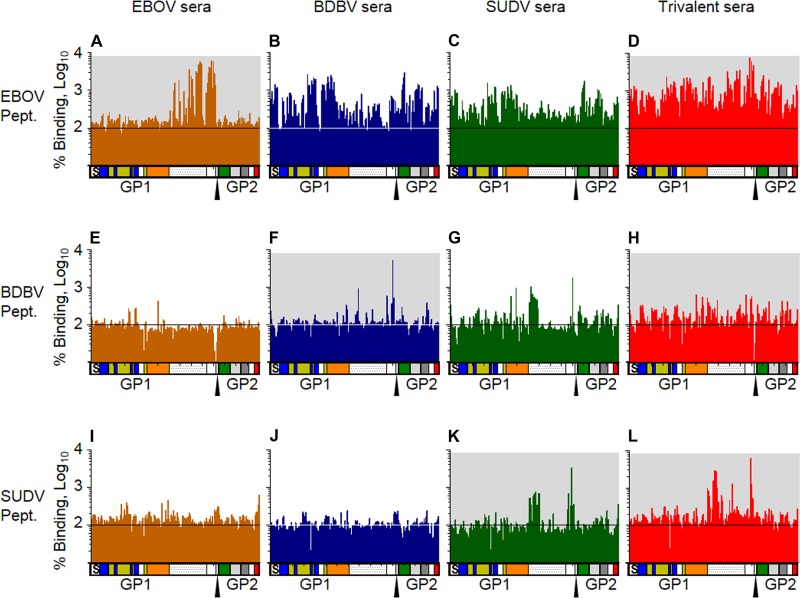
FIG 7.
Trivalent vaccine sera bind GP peptides similarly to single-component vaccine sera. Sera collected at day 56 after immunization with the EBOV vaccine (orange), BDBV vaccine (blue), SUDV vaccine (green), or trivalent vaccine (red) were analyzed for binding to peptides matching the protein sequence of EBOV GP (A to D), BDBV GP (E to H), or SUDV GP (I to L). Values represent the average binding of sera from 3 vaccinated animals as a percentage of the binding of preimmune sera. Each column represents the results for a 15-residue peptide with a sequence matching the sequence of GP. The diagrams below each graph show the domains of GP in a linear fashion that correspond to the binding data. S, signal peptide; blue, base; yellow, head; orange, glycan cap; dotted region, mucin domain; green, fusion loop; light gray, HR1; dark gray, HR2; red, transmembrane and cytoplasmic tail. The furin cleavage site is marked by the triangle. Graphs with a gray background indicate that the vaccine or its component is homologous with the peptide array sequence.

