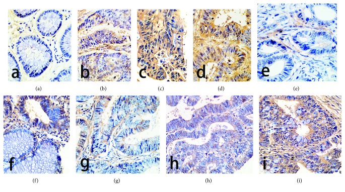
Figure 1.
Epac1 and PDE4 expression in rectal carcinoma tissues (×400). The subfigures (a), (b), (c), and (d) indicated the protein expression of Epac1 in rectal cancer tissues. (a) No expression. (b) Moderately expression. (c, d) High expression levels, expression in the cytoplasm and in the nucleus. The subfigures (e), (f), (g), (h), and (i) indicated the protein expression of PDE4 in rectal cancer tissues. (e) No expression. (f) The top of the picture is moderately expression, and the bottom is no expression. (g) Low expression levels. (h) Moderately expression. (i) High expression levels, mainly in the cytoplasm, with low amounts in the nucleus.

