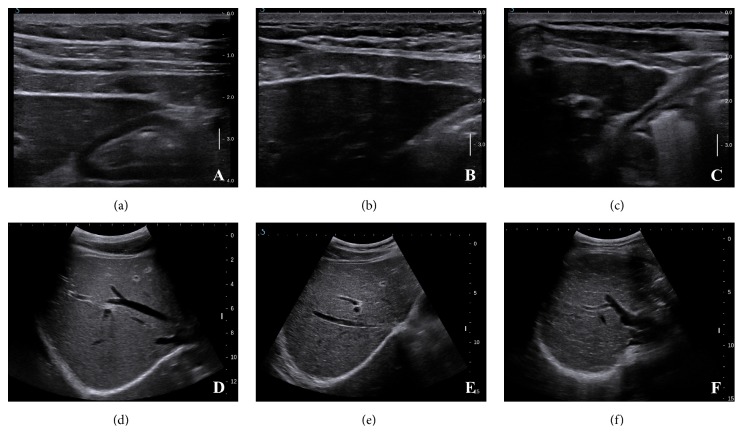
Figure 1.
B-mode images of conventional ultrasonography (US) scoring system. (a) Smooth liver surface, score of 1. (b) Uneven liver surface, score of 2. (c) Irregular nodular liver surface, score of 3. (d) Homogeneous parenchyma, score of 1; and smooth hepatic vein vessel wall, score of 1. (e) Heterogeneous liver parenchyma with fine scattered hyperechoic or hypoechoic areas, score of 2. Obscured or slightly irregular hepatic vein vessel wall, score of 2. (f) Coarse liver parenchyma with an irregular pattern, score of 3.

