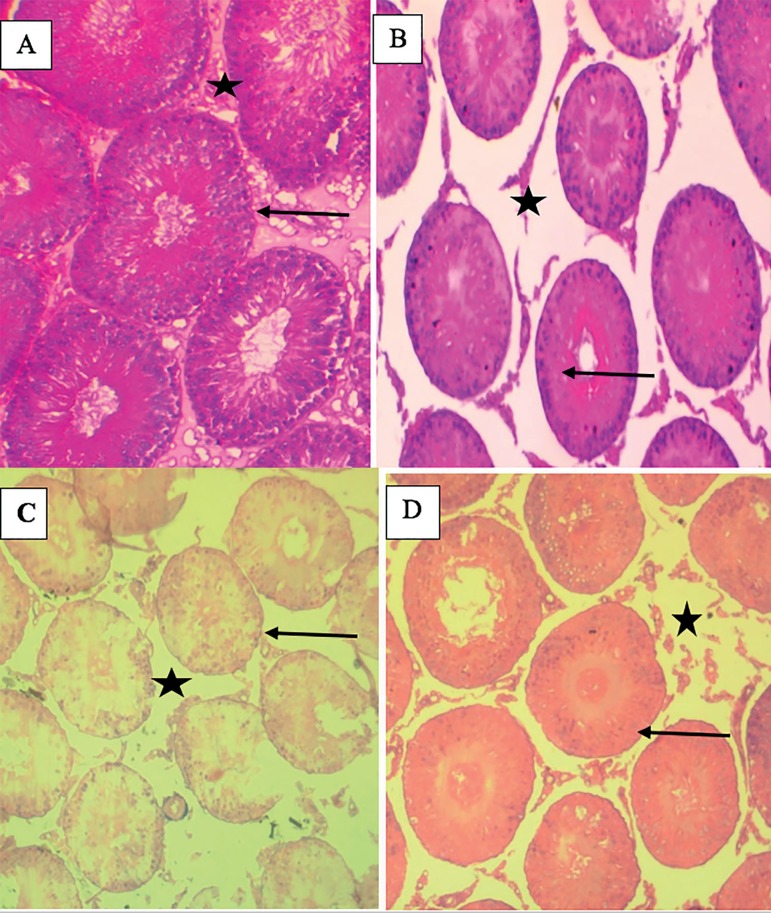
Figure 5.
Photomicrographs of the testes showing seminiferous tubules (black arrow) and their respective interstitial space (black star box) in (A), Control group shows normal seminiferous tubule with complete sperm maturation, normal germinal cells layer, presence of spermatozoa strand in the lumen and interstitial cells appear normal. (B) OG (C) Dia and (D) Dia+OG groups show mild vacuolation in the seminiferous tubule, disorganized germinal cells layer, arrest sperm maturation with empty spermatozoa in lumen. X 100.

