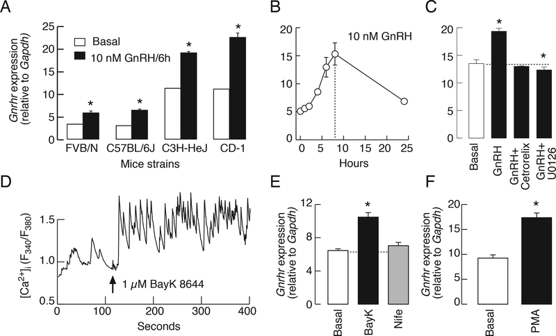
Figure 6.
GnRH also stimulates mouse pituitary Gnrhr expression through calcium and PKC-ERK1/2 signaling pathways. A, Basal and GnRH-stimulated Gnrhr expression in cultured pituitary cells from different mice strains. Asterisks indicate significant differences between pairs. B-F, Cultured pituitary cells from C57BL/6J mice. B, A time course study of GnRH-stimulated Gnrhr expression. C, The lack of effect of 10 nM GnRH on Gnrhr expression in the presence of 1 μM cetrorelix acetate, a GnRHR antagonist, and inhibition of GnRH-stimulated Gnrhr expression by U0126. D and E, Effect of BayK 8644, an L-type voltage-gated calcium channel agonist, on calcium influx (D) and Gnrhr expression (E). Notice the lack of effects of nifedipine—an L-type calcium channel antagonist—on Gnrhr expression. In calcium measurements, the identification of gonadotrophs was done by application of GnRH at the end of recording (not shown). The effects of PMA (100 nM) on Gnrhr expression (F). In all experiments Gnrhr expression was evaluated after 6 h. Experiments were performed in mouse pituitary cells 24 h after dispersion. In A, C, E, and F, asterisks indicate significant differences (P < .05) when compared to basal gene expression, determined by t-test.

