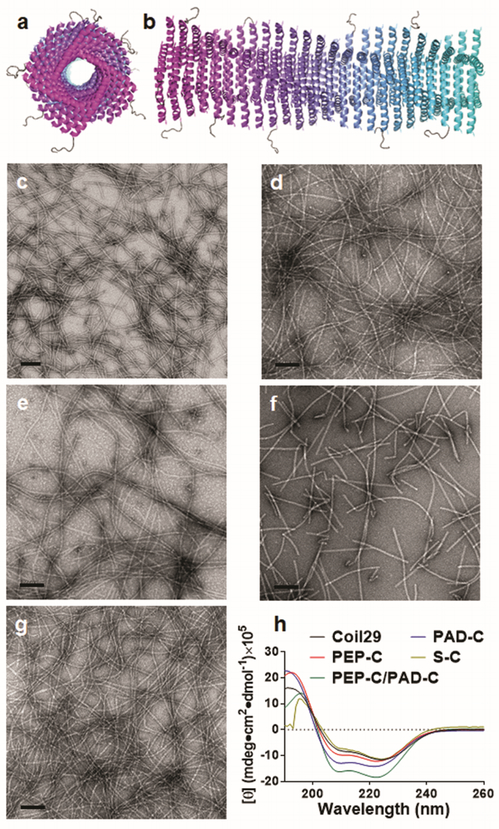
Figure 1.
Coil29 self-assembled into α-helical nanofibers when appended with different epitopes. Axial view (a) and side view (b) schematics of Coil29 fibers displaying PEPvIII epitopes, drawn using PDB structures for Coil29 (PDB ID 3J89) and PEPvIII (PDB ID 1I8I). By TEM, nanofibers were formed by Coil29 (c), PEP-C alone (d), PEP-C and PAD-C co-assembled at 20:1 (e), PAD-C alone (f), and S-C alone (g). Alpha-helical secondary structures were preserved in all fiber formulations as evidenced by circular dichroism spectra (h). All scale bars: 100 nm.

