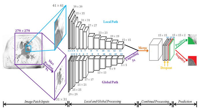
Fig. 1.
The architecture of the proposed dual fully convolutional neural network for atrial segmentation (AtriaNet). The size of the image at every second layer is shown, further details are in Table. I. The parallel (global and local) pathways process each MRI slice at different resolutions, which are combined at the end of the network. The final output has two feature maps, denoting the probability of a positive or negative pixel classification for each 15×15 patch respectively.

