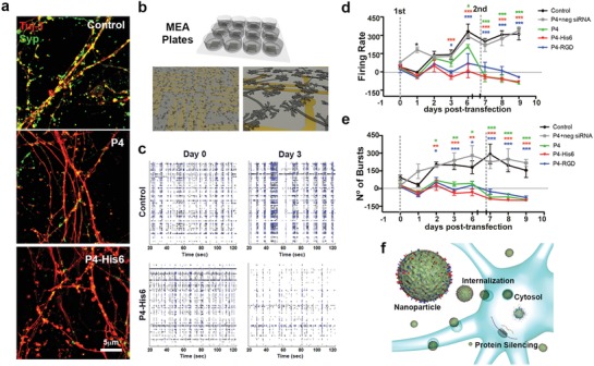
Figure 4.

Electrophysiological studies of functional gene knockdown of neuronal cell cultures by supramolecular particles. a) Structured illumination microscopy (SIM) images of cortical neuronal cells stained with Tuj‐1 (red) and synaptophysin (Syp, green) 24 h after transfection. b) Concept scheme of multielectrode array plates (MEA plates) for neural cell recording. c) Raster plots of a single MEA plate well of representative control and P4–His6‐treated cells before transfection (day 0) and 72 h post‐transfection (day 3). Each line represents the signals detected by a single electrode of the MEA array, during 100 s of recordings. d) Longitudinal progression of spike firing frequency and e) burst number. f) Schematic representation of cells transfected by P4 particles. Two‐way repeated measures one‐way analysis of variance (ANOVA) were performed for firing frequency and burst number. *P = 0.05, **P = 0.001, and ***P = 0.0001. All the experiments were performed using a dose 4 (P4: 104 × 10−6 m, siRNA: 200 × 10−9 m).
