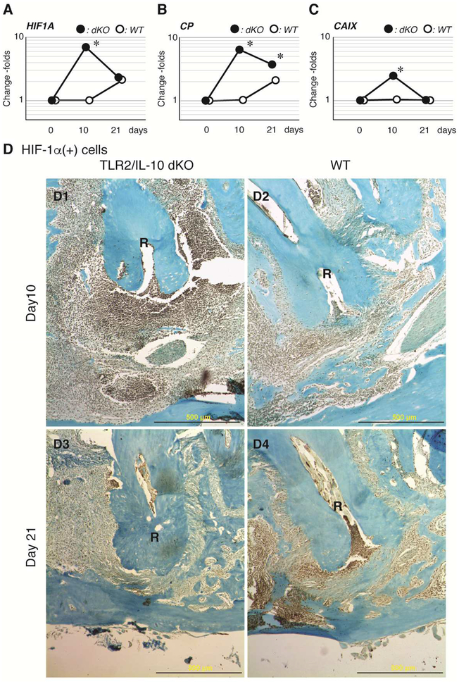
Figure 3. Expression kinetics of HIF-1α in periapical lesions.
qRT-PCR was employed to compare gene expression of Hif1a (A) and its downstream genes Cp (B) and Caix (C) in TLR2/IL-10 KO and WT lesions. Y-axis: logarithmic scale. Closed dot: dKO, Open dot: WT, *: P < 0.05 vs. corresponding day 0 control. (D) HIF-1α protein expression was validated by immunohistochemistry on serial sections of the samples presented in Fig. 1 and 2. (D1) TLR2/IL-10-dKO lesion on day 10 after pulpal infection. (D2) WT lesion on day 10. HIF-1α was faintly expressed in cells around the apical foramen. (D3) TLR2/IL-10-dKO lesion on day 21. (D4) WT lesion on day 21. R: distal root, first molar, Scale bar: 500 μm, 20X magnification.

