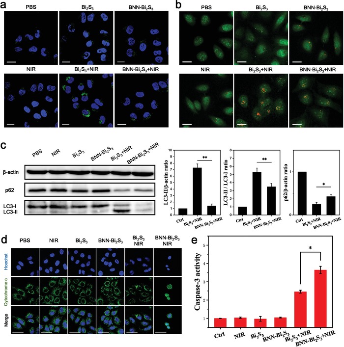
Figure 4.

NO inhibits protective autophagy to sensitize photothermal therapy. a) Confocal microscopy images of BEL‐7402 cells with different treatment. The cells were stained with Hoechst 33342 (blue) and CYTO‐ID Autophagy detection kit (green). b) Confocal microscopy images of BEL‐7402 cells stained with acridine orange (AO). All the scale bars are 20 µm. c) Western blot and its analysis of LC3‐II, LC3‐I, and p62 of BEL‐7402 cells with different treatment. d) Immunofluorescent staining with cytochrome c of BEL‐7402 cells in each group (green). The nuclei were labeled with Hoechst 33342 (blue). The scale bars are 50 µm. e) Relative caspase‐3 activity in BEL‐7402 cells with different treatment. The value of control was set to 1. All the statistical data are shown as mean value and standard deviation, n = 3. p values in (c,e) were calculated by Student's t‐test (** p < 0.01 or *p < 0.05).
