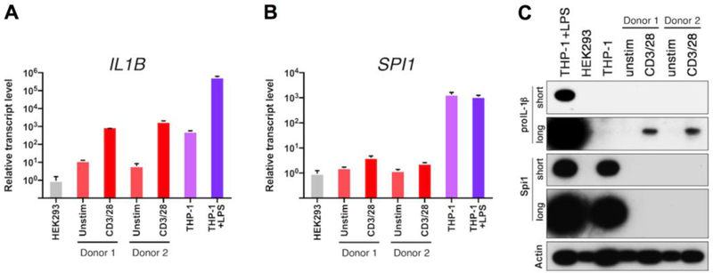
Figure 1. CD4 T cells express proIL-1β following TCR stimulation.
Relative mRNA expression for (A) IL1B and (B) SPI1 in purified CD4 T cells from human lymphoid tissue (two individual patient donors), stimulated for 3 days with anti-CD3/CD28 beads, compared to resting CD4 T cells, HEK293 cells, and THP-1 cells (unstimulated or LPStreated for 1.5 h). Data were normalized to the HEK293 cell sample; standard error represents technical replicates. (C) Western blot analysis of proIL-1β and Spi1 proteins from samples shown in A and B. The proIL-1β and Spi1 blots are shown at short and long exposures to illustrate the relative abundance of these proteins in CD4 T cells. Membranes were stripped and re-probed for β-actin. Note that the β-actin levels vary between cell types and increases with TCR stimulation. Sample inputs for western blots were normalized by cell equivalents: 1.5×10⁵ for all lanes. For conciseness and clarity, only cropped western blot images are displayed. The full-length images of these blots are provided in Supplementary Fig. 3A. Standard error for each donor from lymphocyte populations represents technical replicates, and control cell lines from cultured biologic replicates.

