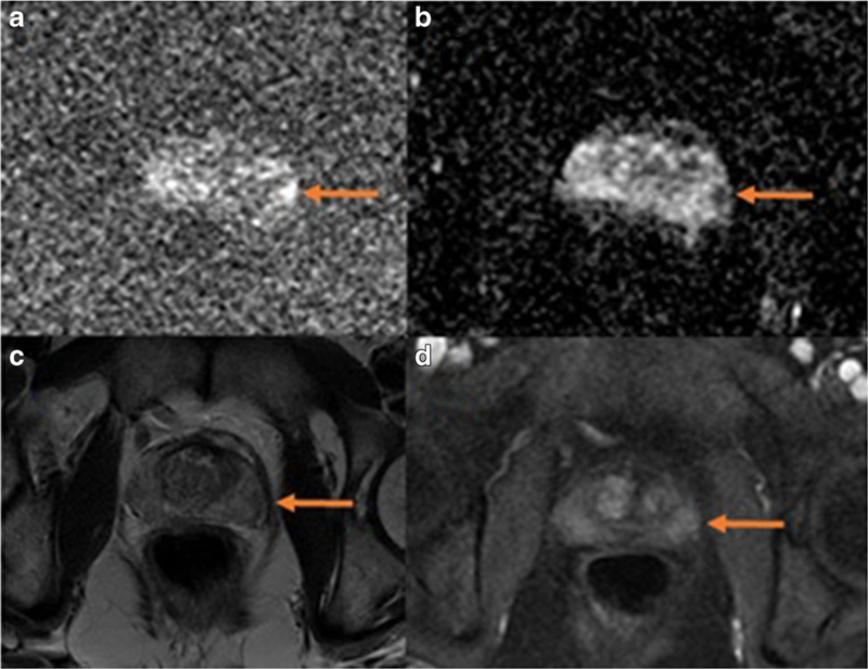
Fig. 1.
A small lesion in the left lateral basal peripheral zone of the prostate gland is seen on mpMRI. The lesion is most conspicuous on DWI as a focus of high signal on high b-value images (a, b=1400) and low signal on ADC map (b). It is barely visible above background on T2-weighted images (c) and DCE (d, early post contrast)

