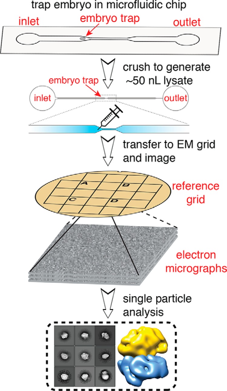
Figure 1.

Schematic of single-cell structural biology approach. Single C. elegans embryos are trapped in a microfluidic device. After the embryo is crushed, the lysate is extracted using a fine needle and applied to a specific area of an EM grid using a stereoscope. The same area is then visualized using EM, and single-particle analysis is applied for structure determination.
