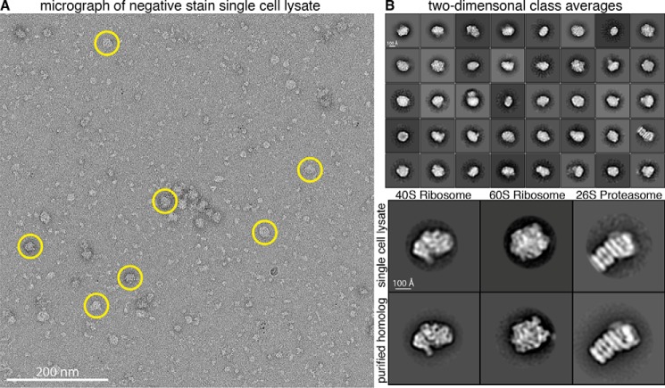
Figure 2.
Single-particle analysis of extracts from single cells. A, representative raw electron micrograph of negatively stained single-cell lysates. Micrographs show monodisperse particles of varying size. Circled particles are representative of the larger particles (∼150–300 Å in diameter) used for subsequent 2D and 3D classification. B, top panel, reference-free 2D alignment and classification of a subset of the ∼50,000 particles picked from single-cell extract. Classes are sorted in order of decreasing abundance. Box size is 576 × 576 Å. Bottom panel, alignment of 2D class averages from single-cell extract to purified homologs.

