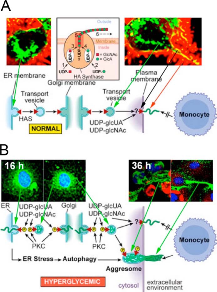
Figure 4.

A, normal glucose: Has enzymes (green) travel to the cell surface, activate hyaluronan synthesis (yellow), and extrude hyaluronan (red) along cell-surface protrusions in nondividing cells. This figure (44) was kindly provided by the Tammi laboratory. B, hyperglycemic glucose: Has enzymes in hyperglycemic dividing cells were activated in intracellular membranes. They then synthesized hyaluronan (green) into ER, Golgi, and transport vesicles after entry into S phase as shown at 16 h of division. After division, they extruded the hyaluronan to form an extracellular hyaluronan matrix as shown at 36 h (16 h after division). Cyclin D3 is localized in intracellular regions (red).
