Figure 6.
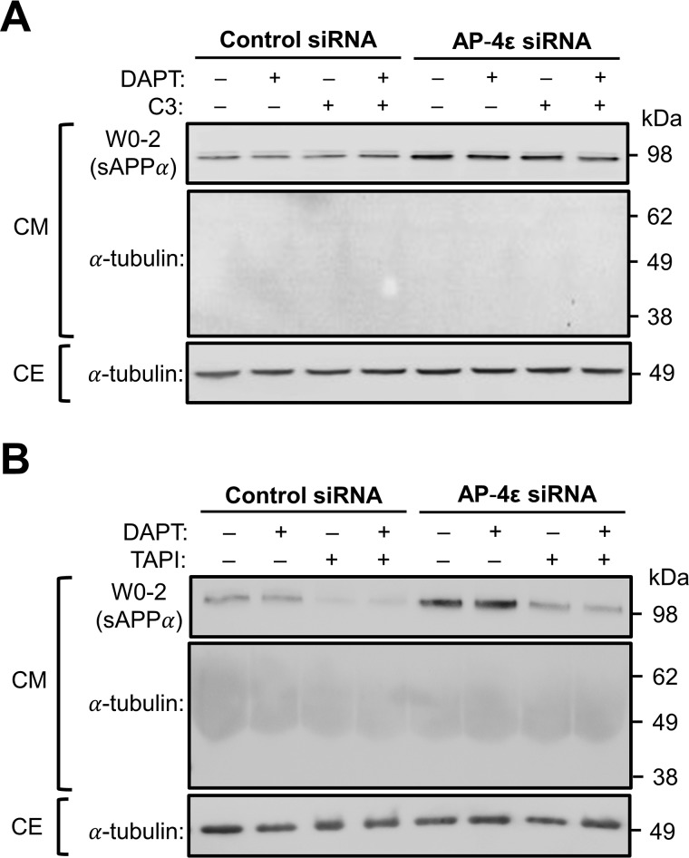
Effect of α-secretase and γ-secretase inhibitors on secretion of the luminal domain of APP. A and B, HeLa–APP695WT cells were transfected with either control siRNA or AP-4ϵ siRNA for 72 h. Cells were also treated with either DMSO carrier (−), 250 nm DAPT (γ-secretase inhibitor), 2 μm C3 (BACE1 inhibitor), or 2 μm C3 and 250 nm DAPT (A) or 50 μm TAPI-1 (α-secretase inhibitor) or 50 μm TAPI-1 and 250 nm DAPT (B) in the last 16–20 h of siRNA transfection. Conditioned medium was collected, and the equivalent volume of samples based on protein content of extracts of cell monolayers were analyzed by immunoblotting with W0-2 antibodies or mouse anti-α-tubulin antibodies. CE were analyzed by immunoblotting with mouse anti-α-tubulin antibodies (the CM collected for analysis in A was carried out in conjunction with the experiment as Fig. 4A; therefore, the immunoblot for α-tubulin in the CE in Fig. 6A is the same as Fig. 4A).

