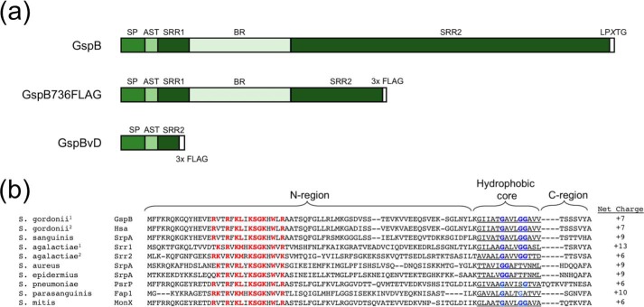
Figure 1.
Domain organization and sequence features of SRR proteins. a, diagram of SRR protein domain architecture, truncated variant GspB736FLAG, and GspB variant D (GspBvD) domains. b, alignment of SRR protein signal sequences of representative Streptococcus and Staphylococcus species. S. gordonii1 is strain M99, S. gordonii2 is strain Challis, S. agalactiae1 is strain COH31, and S. agalactiae2 is strain COH1. Shown are species, protein name, signal sequences, and N-region net charge. Red, residues of the conserved polybasic motif. Blue, conserved glycine residues. Highlighted are the predicted N-region, hydrophobic core, and C-regions of the signal peptide sequences.

