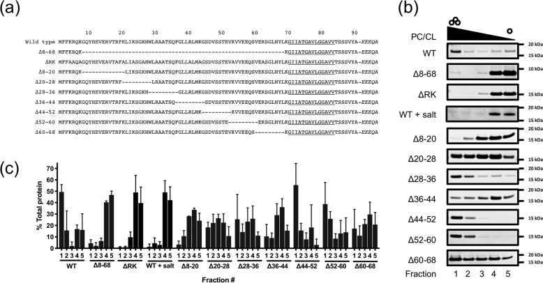
Figure 3.
GspB lipid binding is mediated by the N-region. a, diagram of signal peptide N-region mutants studied here. Italics denote residues in the mature region of GspB. b, comparison of lipid binding of GspB signal peptide variants with PC/CL liposomes. Representative Coomassie-stained SDS-PAGE gels are shown. c, densitometric quantification of signal peptides in gradient fractions from b. Independent experiments were performed in triplicate. Plotted are mean values ± S.D. (error bars).

