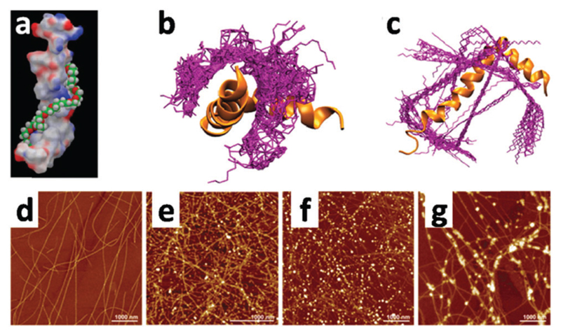
Fig. 21.
(a) Atomistic model of a PEG chain docked to Aβ1–42. The chain interacts with both hydrophobic as well as hydrophilic residues of the peptide and forms a spiral structure. (b) Best 50 conformations of the PEG chain (purple) docked to Aβ1–42 (orange), and (c) alkyl PE chain (purple) docking on Aβ1–42 (orange). Images (a–c) are reproduced with permission from ref. 368. Copyright 2012, American Chemical Society. (d–g) AFM (bottom row) images of the β-lactoglobulin amyloid fibrils (d) and fibrils decorated by gold (e), silver (f) and palladium (g) nanoparticles after the respective metal salt reduction by NaBH4. Images (d)–(g) are reproduced with permission from ref. 370. Copyright 2014, American Chemical Society.

