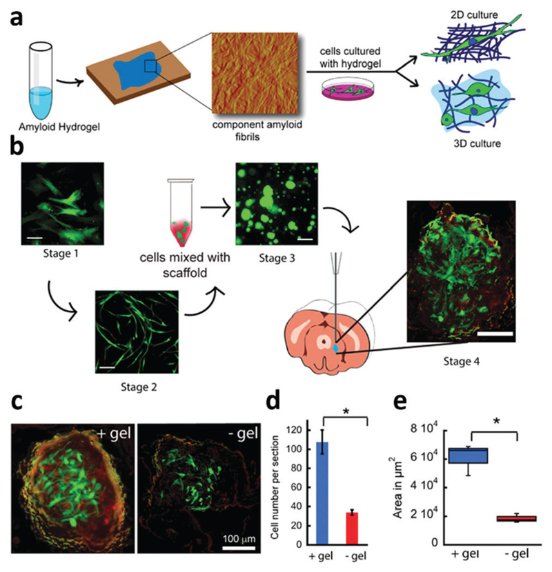
Fig. 29.
Amyloid hydrogels for cell culture: (a) schematic of amyloid hydrogels for 2D and 3D cell cultures. (b) Schematic of the morphology of cells at each stage during the implantation. Stage 1: cultured cells for 24 h; stage 2: cells were primed with a differentiation medium for 5 days; stage 3: cells were then transplanted with hydrogel A5 into the mice brains, and stage 4: the brains were harvested. Scale bars: 200 μm for stage 1–3 and 100 μm for stage 4. (c) Implanted GFP-hMSCs with α-synuclein hydrogel (left) and without a hydrogel (right) at the caudate putamen after 7 days in vivo. (d) Cell viability when implanted with and without a hydrogel. (e) Box plot of the area with survived cells when transplanted with and without a hydrogel. Reproduced with permission from ref. 422. Copyright 2016, Nature Publishing Group.

