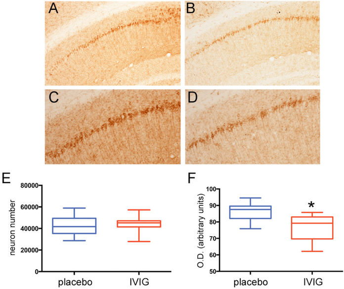
Fig. (1). Three months of IVIG reduces NFT expression levels within CA1 neurons. A-D).
Photomicrographs show AT-180 immunolabeling of CA1 pyramidal neurons and fibers in 3xTg mice treated with placebo (A,C) or IVIG (B,D) for three months. Panels (C) and (D) are higher magnification photomicrographs of (A) and (B), respectively. Note the lighter staining in the IVIG-treated mice compared to placebo-treated mice. E) Unbiased stereologic cell counts revealed no difference in the number of AT-180+ CA1 neurons between the two groups. F) Optical densitometric measurements revealed a significant 15–20% decrease in AT-180 labeling intensity within individual CA1 neurons in IVIG compared to placebo. n = 12/group; *, p< 0.01 via Student’s unpaired t test (two-tailed).

