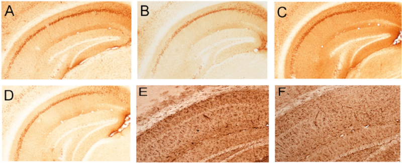
Fig. (4). IVIG effects on hippocampal intracellular amyloid and astroglial pathology.
A-D) Photomicrographs show immunolabeling of CA1 neurons and fibers with the 6E10 antibody raised against the N-terminus of Abeta. Intraneuronal amyloid accumulation was observed following three months of placebo (A) or IVIG (B) treatment. Note the relatively lighter cellular and neuropilstaining in the IVIG treated group. Intraneuronal Abeta continued to accumulate following six months of placebo (C) treatment but remained diminished with IVIG (D) treatment. E,F) Photomicrographs show GFAP immunolabeling of astroglia in the hippocampal CA1 region in the placebo (E) and IVIG (F) groups after three months treatment. Note the relatively lighter staining in IVIG compared to placebo. This differential staining pattern continued in the six month treatment groups (not shown).

