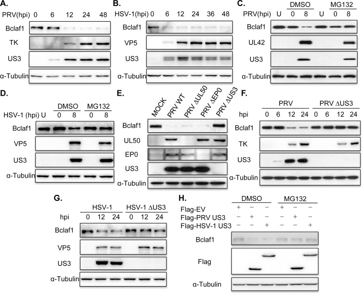
Fig 1. PRV and HSV-1 employ US3 to decrease Bclaf1 in a proteasome-dependent manner.
(A) IB analysis of Bclaf1, TK and US3 in PK15 cells infected with PRV (MOI = 1) for the indicated hours. α-Tubulin was used as loading control. (B) IB analysis of Bclaf1, VP5 and US3 in HEp-2 cells infected with HSV-1 (MOI = 5) for the indicated hours. (C) IB analysis of Bclaf1, UL42 and US3 in PK15 cells infected with PRV (MOI = 1) followed by untreatment (U) or treatment with DMSO or MG132. (D) IB analysis of Bclaf1, VP5 and US3 in HEp-2 cells infected with HSV-1 (MOI = 5) followed by untreatment (U) or treatment with DMSO or MG132. (E) IB analysis of Bclaf1, UL50, EP0 and US3 in PK15 cells infected with indicated PRV strains (MOI = 1) at 8 hpi. (F) IB analysis of Bclaf1, TK and US3 in PK15 cells infected with PRV WT or PRV ΔUS3 (MOI = 1) for the indicated hours. (G) IB analysis of Bclaf1, VP5 and US3 in HEp-2 cells infected with HSV-1 WT or HSV-1 ΔUS3 (MOI = 5) for the indicated hours. (H) IB analysis of endogenous Bclaf1 in HEK293T cells transfected with Flag-tagged PRV/HSV-1 US3 expression plasmids followed by treatment with DMSO or MG132.

