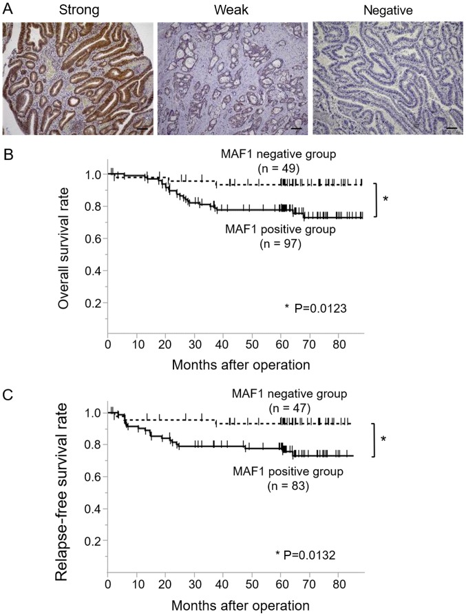
Figure 1.
Immunohistochemical analysis of MAF1 expression and its prognostic impact in CRC. (A) Representative cases of immunohistochemical staining with anti-MAF1 antibody. Examples of strong intensity, weak intensity and negative staining are presented. Scale bar, 100 µm. Negative staining was defined as the MAF1-negative group, whereas strong and weak staining was defined as the MAF1-positive group. Kaplan-Meier curves for (B) overall survival and (C) recurrence-free survival, according to MAF1 expression status in patients with CRC. CRC, colorectal cancer; MAF1, MAF1 homolog, negative regulator of RNA polymerase III.

