Figure 2.
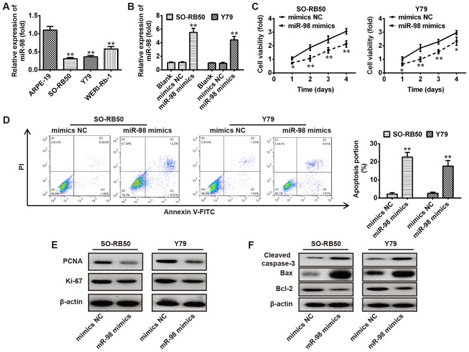
Overexpression of miR-98 suppresses RB cell growth. (A) RT-qPCR was conducted to determine the expression levels of miR-98 in human RB cell lines, including WERI-Rb-1, Y79 and SO-RB50, and normal retinal pigmented epithelium cells, ARPE-19. (B) miR-98 expression levels in SO-RB50 and Y79 cells following transfection with miR-98 mimics or NC were measured via RT-qPCR. (C) An MTT assay was used to analyze the cell viability of SO-RB50 and Y79 cells transfected with miR-98 mimics or NC. (D) Cell apoptosis was measured via flow cytometry analysis in SO-RB50 and Y79 cells transfected with miR-98 mimics or NC. (E and F) SO-RB50 and Y79 cells were transfected as aforementioned (C), and western blot analysis was performed to detect the expression of proliferation markers (PCNA and Ki-67) and apoptosis-associated proteins (cleaved-caspase-3, Bax and Bcl-2) in SO-RB50 and Y79 cells, respectively. β-actin was used as an internal control for protein loading. Data are presented as the mean ± standard deviation of three independent experiments. *P<0.05, **P<0.01 vs. ARPE-19 cells, blank or NC. Bcl-2, B cell lymphoma-2; Bax, Bcl-2-associated X; FITC, fluorescein isothiocyanate; miR, microRNA; NC, negative control; PCNA, proliferating cell nuclear antigen; PI, propidium iodide.

