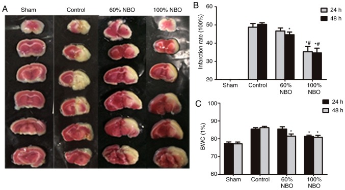
Figure 1.
Quantification of infraction rate and brain water content at 24 and 48 h. (A) Brain sections stained with 2,3,5-triphenyltetrazolium chloride exhibiting ischemic infarctions. Red-colored regions indicate non-ischemia, and pale-colored regions indicate the ischemic portion of the brain. (B) Infraction rate at 24 and 48 h. (C) BWC at 24 and 48 h. *P<0.05 vs. control group; #P<0.05 vs. 60% NBO group. Values present the mean ± standard deviation, n=3 per group. BWC, brain water content; NBO, normobaric oxygen.

