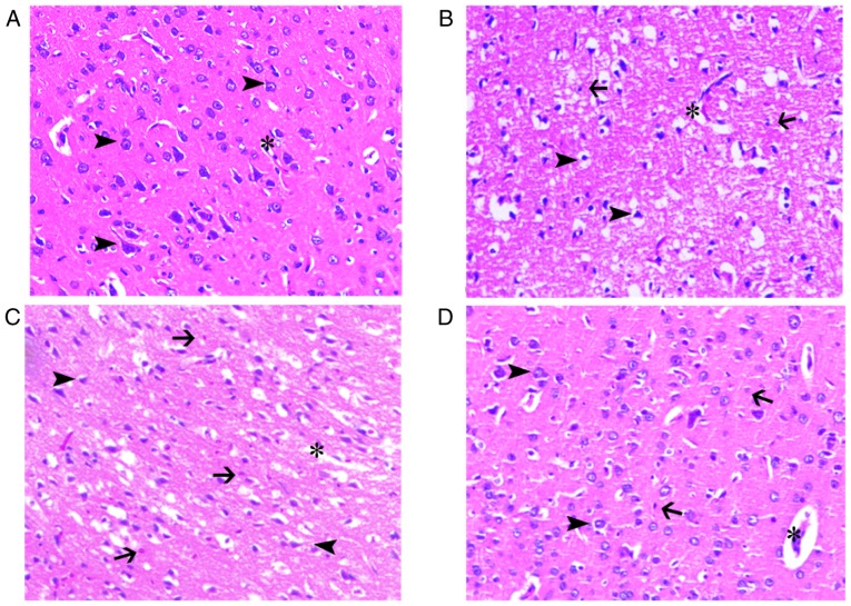
Figure 2.
Histological were evaluated by haematoxylin and eosin staining of cerebral ischemic penumbra at 48 h. Morphological improvement in slice following NBO treatment. (A) The normal brain tissue was depicted. (B) The control group numerous shrunken and triangular-shaped neurons with nuclei shrinkage distributed in the damaged brain tissue brain edema, neutrophil infiltration and increased perivascular space were also observed. (C) Brain tissue damage exhibited less damage in the 60% NBO group, compared with the vehicle control. (D) The 100% NBO treatment ameliorated brain damage, compare with 60% NBO treatment. x200 magnification. Arrow head, neurons; asterisk, capillaries; arrow, neutrophils; NBO, normobaric oxygen.

