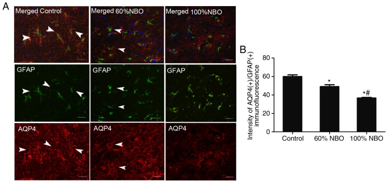
Figure 6.
Characterization of AQP4 expression in the astrocytic compartment of the control and NBO treatment rat brains at 48 h. Triple immunofluorescent staining of GFAP (green), AQP4 (40) and DAPI (blue). (A) The immunofluorescence of AQP4(+)/GFAP(+) expression in the control and NBO treatment groups. (B) The quantification of AQP4(+)/GFAP(+) immunofluorescence in the control and NBO treatment rats group (3-5 fields was observed). *P<0.05 vs. the control group; #P<0.001 vs. the 60% NBO group. Scale bars, 50 µm. Arrow head, co-expression of AQP4(+)/GFAP(+) astrocytes; NBO, normobaric oxygen; AQP4, aquaporin-4; GFAP, glial fibrillary acidic protein.

