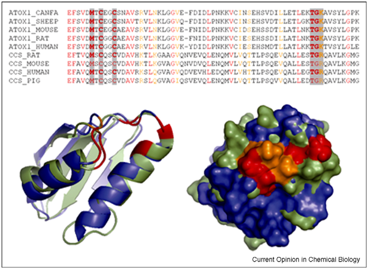
Figure 2.
Structural comparison of human Atox1 (blue, 1TL5) and Domain I of CCS (green, 2CRL). Sequence alignment illustrates significant similarity of mammalian Atox1 and CCS (identical residues are in red, conserved residues in orange). The conserved residues around the metal-binding site are highlighted by grey (top); their location in the superimposed structures (left) and surface exposure (right) are shown (bottom).

