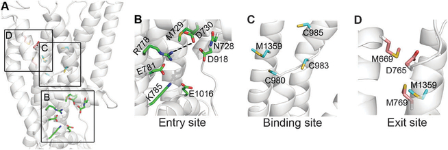
Fig. 5. Potential functional sites in the membrane region.
The ATP7B model is shown as transparent white cartoons, and viewed from the side with the inward side facing below. (A) Focus on the membrane domain, with residues of the functional site (Table 1) shown as colored sticks. The three sites, i.e. entry, intra-membrane binding and exit sites, are marked by squares. (B) The entry site, with suggested functional residues as green sticks. R778 and D730 could be salt-bridged. (C) The binding site, with TM3 removed for clarity. The suggested binding site residues (colored cyan) are C983, C985 and Met1359 with C980 playing an assisting role. (D) Exit site residues in pink, along with Met1359. TM2 and TM3 were extracted for clarity.

