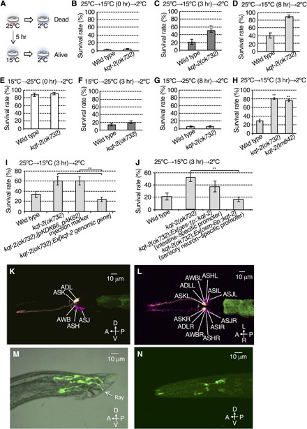
Fig. 1. KQT-2 in sensory neurons regulates cold acclimation.

(A) Cold acclimation assay. Wild-type animals cultivated at 25°C fail to survive at 2°C, whereas animals cultivated at 15°C survive at 2°C. When wild-type animals cultivated at 25°C are transferred to and conditioned at 15°C for 5 hours, they exhibit increased survival at 2°C. (B to D) Cold acclimation of kqt-2(ok732) mutants assayed by the 25°C→15°C→2°C protocol. kqt-2(ok732) exhibited supranormal cold acclimation. Number of assays ≥ 12. (E to G) Cold acclimation of kqt-2(ok732) mutants assayed by the 15°C→25°C→2°C protocol. Number of assays ≥ 10. (B to G) Error bar indicates SEM. Comparisons were performed using the unpaired t test (Welch). *P < 0.05, **P < 0.01. (H) Two kqt-2 loss-of-function mutants exhibited supranormal cold acclimation. Number of assays ≥ 11. Error bar indicates SEM. Comparisons were performed using Dunnett’s test. *P < 0.05, **P < 0.01. (I) Transgenic rescue of kqt-2(ok732) with a genomic fragment encompassing the wild-type kqt-2(+) gene and native promoter sequence. Number of assays ≥ 8. (J) Rescue of kqt-2 mutants by tissue-specific expression of kqt-2 cDNA. Number of assays ≥ 9. (I and J) Error bar indicates SEM. Comparisons were performed using the Tukey-Kramer method. *P < 0.05, **P < 0.01. (K to N) kqt-2::GFP driven by the kqt-2 promoter is expressed in ADL and ASK head sensory neurons. GFP fluorescence of ADL and ASK (yellow) neurons are colabeled with DiI, which labels only six pairs of amphid sensory neurons in the head with red fluorescence (magenta). (K) Lateral view. A, anterior; P, posterior; D, dorsal; V, ventral. (L) Ventral view. L, left; R, right. (M) GFP driven by the kqt-2 promoter is expressed in fan and ray sensory neurons in the tail of males. (N) Wild-type animal expressing kqt-2cDNA::gfp (8 ng/μl) using a 9.0-kb kqt-2 promoter. KQT-2::GFP is observed in whole sensory neurons and is especially localized to sensory endings and cell bodies of head sensory neurons.
