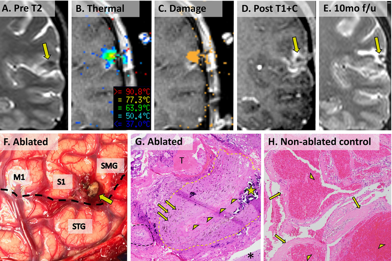
Figure 3. SLA and eventual resection of CCM in patient 16 with pathological comparison to an non-ablated CCM from an unrelated patient.
(A) Preoperative coronal T2 image showing CCM just above the Sylvian fissure in the postcentral sulcus. (B) Visualase ablation screenshot showing thermal temperature map. (C) Visualase ablation screenshot showing cumulative irreversible damage estimate (orange pixels). (D) Contrasted T1 image showing final extent of ablation. (E) Delayed post-ablation T2 image showing smaller hypointense CCM with surrounding hyperintense ablation cavity. (F) Operative photograph of ablated CCM (tan nodule, arrow) posterior to precentral gyrus (M1) between postcentral gyrus (S1) and supramarginal gyrus (SMG) and superior to the Sylvian fissure (dotted black line) and superior temporal gyrus (STG). (G) 100x microscopic appearance of patient 16’s CCM (H&E stain) showing post-ablation reactive type changes surrounding a large collapsed thickened vessel (perimeter approximated by dotted yellow outline and collapsed lumen lined by arrowheads). Scattered prior hemorrhage (arrows) and calcification (star) are evident within the thickened vessel wall. A small sclerotic hyalinized vessel (dotted black outline), an acute extravascular/extraparenchymal thrombus (T, likely a surgical artifact), and reactive brain parenchyma (asterisk) surround the collapsed vessel. (H) For comparison, a surgically resected CCM from an unrelated patient shows multiple engorged, dilated vascular lumina (arrowheads) with thickened, hyalinized walls (arrows) without intervening brain parenchyma.

