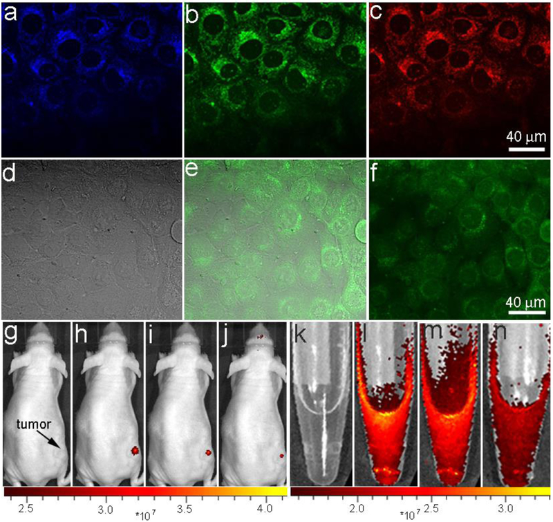
Fig. 3.
In vitro and in vivo FL imaging. Laser scanning confocal microscopy images of SF-763 cells incubated with BFNPs for 2 h under different excitation wavelengths: (a) 405 nm; (b) 488 nm; (c) 546 nm. (d) Transmission, (e) two-photon fluorescence and (f) overlaid images of SF-763 cells incubated with BFNPs. Excitation laser wavelength was 820 nm. NIR FL images of BFNPs injected into a tumor (g-j) and BFNPs (k-n) in PBS solution (0.5 mg/mL, inset) under various excitation wavelengths: (g and k) white light, (h and l) 605 nm, (i and m) 640 nm and (j and n) 675 nm.

