Figure 1:

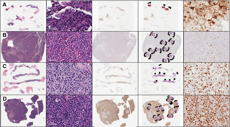
Images from representative cases showing (row A) insufficient material for review, (row B) negative MYC immunohistochemical (IHC) staining, (row C) equivocal MYC IHC staining (percent positivity in 30-50% range), and (row D) positive MYC IHC staining. The first column shows whole-slide images of the hematoxylin and eosin (H&E) stained slide. The second column shows a portion of the H&E slide at 400x magnification, in one of the selected areas of best tissue. The third column demonstrates whole-slide images of the MYC IHC slide. The fourth column shows the Aperio ImageScope annotations on the MYC IHC slide, with black marking pen indicating the areas of best quality tissue selected as the areas to evaluate. The red boxes select tissue to analyze, while the text and ruler icons in blue measure the area selected, with the goal of 1 mm2 in each area. The fifth column shows the MYC IHC slide at 400x magnification, within one of the evaluation areas.
