Figure 1.
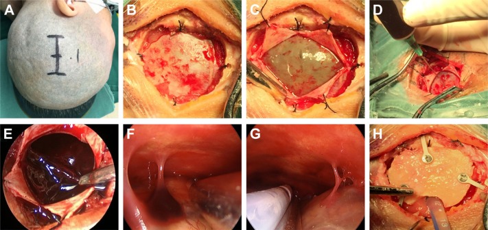
The basic steps of neuroendoscopic hematoma evacuation for chronic and subacute subdural hematoma.
Notes: (A) The patient was placed in the lateral position, and a straight scalp incision was made on the parietal region. (B) After a bone flap was created, the dura was suspended. (C) After the dura was opened in a cruciform fashion, the outer membrane of the hematoma was revealed. (D) The liquefied blood was slowly aspirated with a syringe to decrease the intracerebral pressure. (E) The residual hematoma and blood clots were evacuated by suction under the neuroendoscope. (F) Fibrin septa were checked and treated under the neuroendoscope. (G) A bridging vein was seen, and a draining catheter was introduced in the cavity under the neuroendoscope. (H) The bone flap was replaced and fixed.

