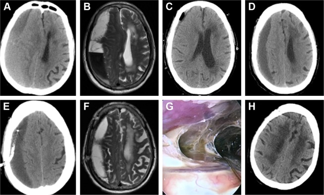
Figure 3.
Neuroendoscopic surgery for recurrent CSDH.
Notes: (A) CT scan showing a CSDH on the right side. (B) MRI showing fibrin septa in the hematoma cavity. (C) CT scan showing the incomplete evacuation of the hematoma one day after the first burr-hole surgery. (D) CT scan showing hematoma reaccumulation 10 days after the first burr-hole surgery. (E) CT scan showing the hematoma in the subdural area 1 day after the second burr-hole surgery. (F) MRI showing that the hematoma had not disappeared 15 days after the second burr-hole surgery. (G) Operation view showing fibrin septa in the hematoma cavity during the third surgery via the neuroendoscope. (H) Forty days after the third surgery by neuroendoscope shows no recurrence.
Abbreviations: CSDH, chronic subdural hematoma; CT, computed tomography; MRI, magnetic resonance imaging.

