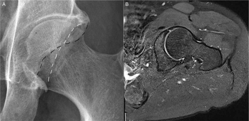
Figure 5.

(A) AP view of the right hip. The anterior (white dots) and posterior (black dots) rim of the acetabulum are marked. The superior portion of the anterior rim lies lateral to the posterior rim indicating overcoverage of the acetabulum. Anteriorly, it assumes a more normal medial position, creating the crossover sign as a positive indicator of pincer impingement. (B) Fat-suppressed oblique axial proton density weighted image shows linear high signal separating the anterior labrum roughly in halves, a radial tear.
