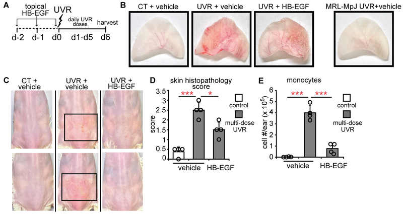
Fig. 8. Topical EGFR ligand reduces photosensitivity.
(A) Experimental scheme for (B-E) (n= 4 mice). MRL-Faslpr mice ears and back skin were topically treated with HB-EGF for 2 days before and on the first day of UVR exposure and examined 24 hours after the final exposure. (B) Representative images of ears. The MRL-MpJ ear represents a non-SLE control. (C) Representative images of back skin; boxes outline lesional areas. Magnified images of back skin in Fig. S11. (D) Ear histopathology score. (E) Absolute monocyte numbers. (B-E) Data are from 3 independent experiments. Bars represent means. Error bars depict standard deviations. *p<0.05, ***p<0.001 using two-tailed unpaired Student’s t-test after one-way ANOVA.

