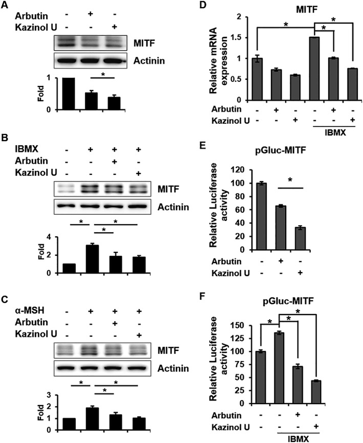
Figure 3.

Inhibition of MITF mRNA, protein and transcriptional activity by kazinol U. (A) Cells were treated with arbutin (500 μM), kazinol U (20 μM) or DMSO for 6 h. (B–D) Cells were pretreated with arbutin, kazinol U or DMSO for 1 h and then cultured with IBMX (100 μM) or α‐MSH (1 μM) for 6 h (B, C) or 24 h (D). (A–C) MITF expression was detected by Western blot analysis (A: n = 5, B and C: n = 7). (D) MITF mRNA expression was measured by quantitative RT‐PCR using specific primers (n = 5). (E, F) Cells were co‐transfected with pMITF‐Gluc and a β‐galactosidase plasmid. The treatment condition was the same as in Figure 2. The changes in luciferase activity with respect to the DMSO‐control were calculated (E and F: n = 6). One‐way ANOVA with a post hoc Newman–Keuls test was used for the statistical analysis; *P < 0.05.
