Figure 2.

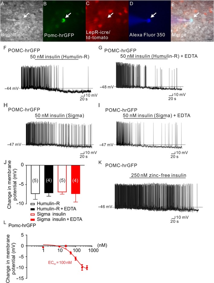
Insulin hyperpolarizes arcuate POMC neurons independent of zinc. (A–E) Brightfield illumination (A) of a POMC neuron that does not express leptin receptors from POMC‐hrGFP::LepR‐cre::td‐tomato mice. (B) and (C) show the same neuron under FITC (hrGFP, green cell) and Alexa Fluor 594 (td‐tomato, not red cell) illumination. Complete dialysis of Alexa Fluor 350 from the intracellular pipette is shown in (D) and merged image of a POMC neuron targeted for electrophysiological recording (E). (Arrow indicates the targeted cell. Scale bar = 50 μm). (F,H) Representative traces depicting a POMC neuron hyperpolarized by insulin (50 nM, Humulin‐R; F) and insulin (50 nM, Sigma insulin; H). (G,I) Administration of EDTA does not block either the Humulin‐R (50 nM) or the Sigma insulin (50 nM) induced hyperpolarization of POMC neurons. (J) Histogram illustrates the acute effects of insulin on the membrane potential of POMC neurons with or without EDTA. Error bars indicate SEM. (Humulin‐R: n = 6; Humulin‐R + EDTA: n = 7; Sigma insulin: n = 7; Sigma insulin: n = 7. Data from 11 mice.) (K) Representative traces showing that POMC neurons are hyperpolarized by zinc‐free insulin (250 nM). (L) Dose‐response of zinc‐free insulin on POMC neurons. (n = 41. Data from 13 mice.)
