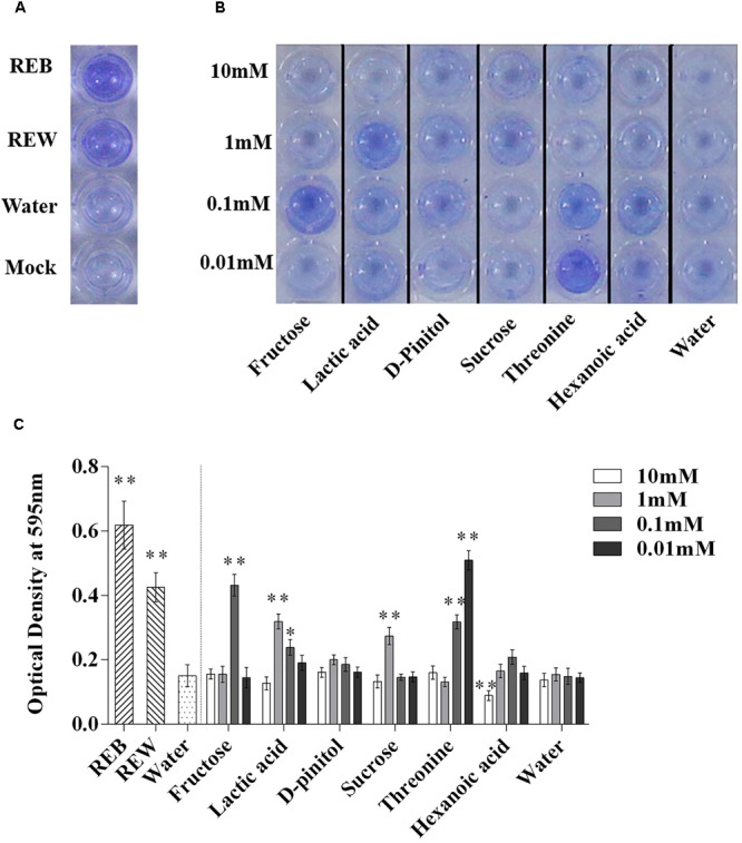
FIGURE 8.

Biofilm formation on polystyrene microtiter plates by Bacillus cereus AR156 with still at 30°C for 48 h under microaerobic conditions. Biofilm formation were determined by crystal violetin in the 96-well microtiter plate induced by Bacillus cereus AR156 (REB), tomato root exudate induced by water (REW) (A) or different components from tomato root exudates induced by B. cereus AR156 (B). (C) Relative optical density of B. cereus AR156’s biofilm was measured at 595 nm with a microtiter plate reader. The mean and standard error values of three biological replicates are reported for each treatment. Asterisks in (C) indicate statistical significance difference between data of treatments and data of sterile water as determined by the Student’s t-test (∗P < 0.05, ∗∗P < 0.01). The experiment was carried out three times and one representative experiment is reported.
