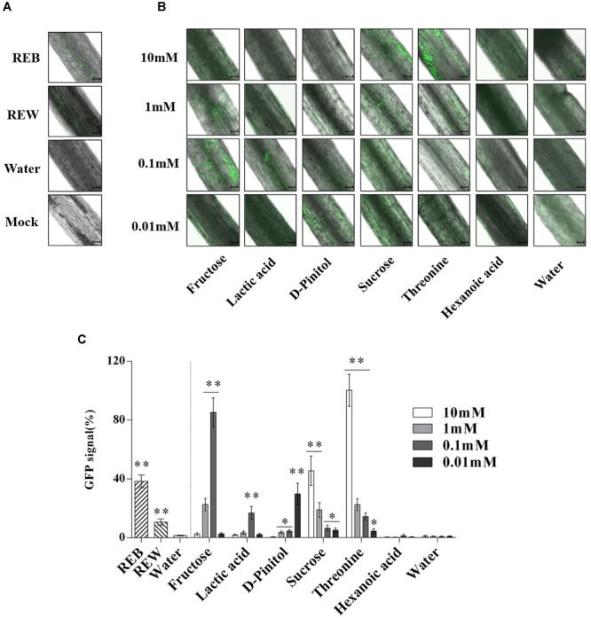
FIGURE 9.

Attachment of GFP-targeted Bacillus cereus AR156 strain on tomato root surface. Cells of Bacillus cereus AR156 form different colonization on the surfaces of excised tomato roots in 1% MS liquid medium containing tomato root exudate induced by Bacillus cereus AR156 (REB), tomato root exudate induced by water (REW) (A) or different components from tomato root exudates induced by B. cereus AR156 (B) at 25°C for 3 days and were visualized by Confocal Laser Scanning Microscope. Mock showed tomato root grown in absence of B. cereus AR156 cells. Scale bars = 100 μm. The results in (A,B) were quantified in (C). GFP signal was quantified using the Confocal Laser Scanning Microscope software (Leica AF6000 Modular microsystems) by measuring the total GFP fluorescence in one field inside the infiltration area with a low magnification objective (20X); all images used for fluorescence measurement were taken with the same settings. Basal signal measured in mock was subtracted from the values measured for each experimental condition, and the signal obtained with 10 mM Threonine was set as 100%. The mean and standard error values of three biological replicates are reported for each treatment. Asterisks in (C) indicate statistical significance difference between data of treatments and data of sterile water as determined by the Student’s t-test (∗P < 0.05, ∗∗P < 0.01). The experiment was carried out three times and one representative experiment is reported.
