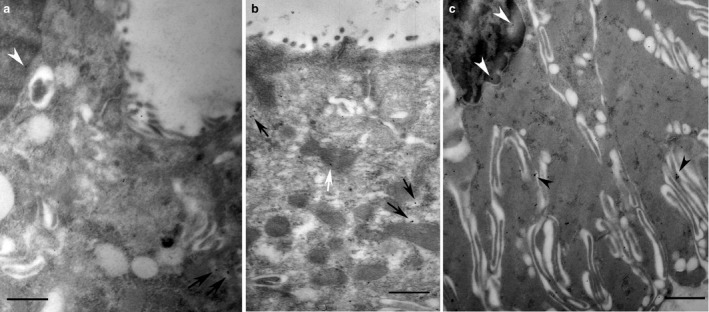
Figure 2.

Intercalated (a) and striated (b,c) duct cells of non‐stimulated parotid glands used for the immunogold localisation of melatonin. The secretory granules of both intercalated and striated ducts displayed no immunoreactivity for melatonin. The small vesicles diffused in the cytoplasm appear to be, however, labelled (black arrows). In striated duct cells (b) melatonin labelling seemed to be also in the folds (arrowheads) of basal membranes and in mitochondria (white arrow). Often, in both ducts, melatonin reactivity occurred in the nuclei (a,c). Scale bar: 1 μm.
