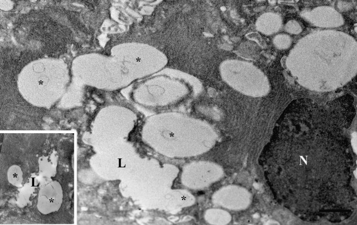
Figure 3.

Cells of parotid gland by transmission electron microscopy (TEM) exposed to isoprenaline for 60 min post‐fixed with osmium to enhance morphology. The lumina (L) were dilated and profiles of exocytosis (asterisks) were visible. N, nucleus. Insert: Omega exocytotic profiles clearly connected to the lumen. Scale bar: 1 μm.
