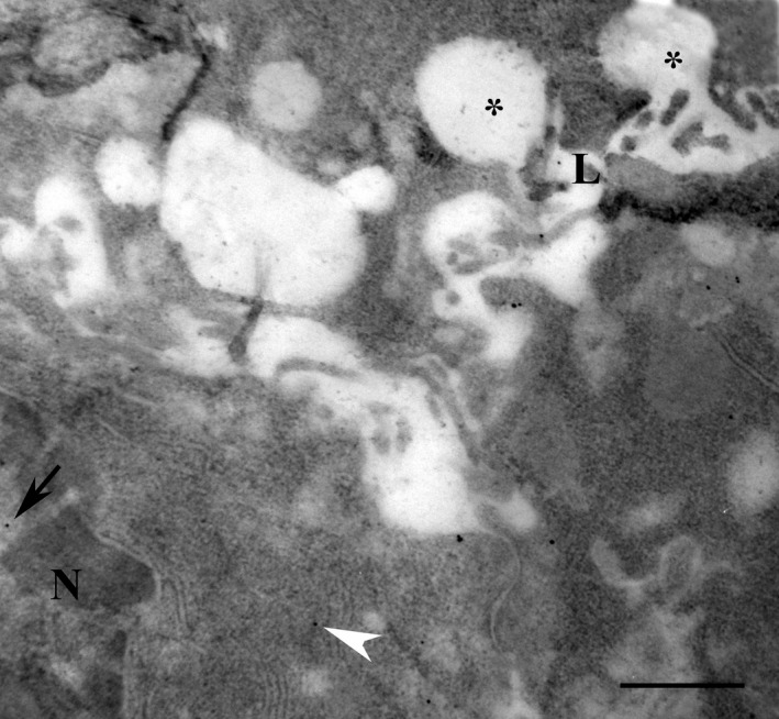
Figure 4.

Serous cells of parotid gland exposed to isoprenaline for 60 min and used for the immunogold localisation of melatonin. Melatonin reactivity was observed in the lumen (close to the letter L) and in the membranes (white arrowhead) as well as in the nucleus (arrow). N: nucleus. *Omega exocytosis profiles. Scale bar: 1 μm.
