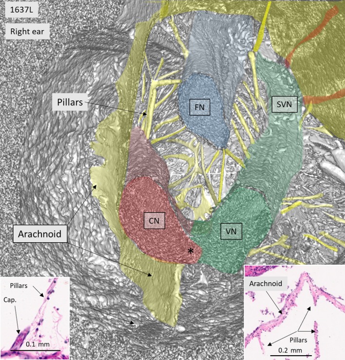
Figure 3.

SR‐PCI and 3D reconstruction of IAC shown in Fig. 2, cross‐sectioned more medially. The cranial nerves with radiating pillars can be seen. Left and right insets represent H&E‐stained celloidin sections of a cross‐sectioned human IAC. The cistern wall consists of an arachnoid membrane surrounding the nerve complex. Pillars run between the nerves and the arachnoid and are occasionally associated with capillary vessels (left inset). Both the arachnoid and pillars consist of connective tissue fibers surrounded by fibrous cells. Histological sections were kindly provided by Dr Charles Liebermann, Massachusetts Eye and Ear Infirmary, Boston, MA, USA.
